Views: 6



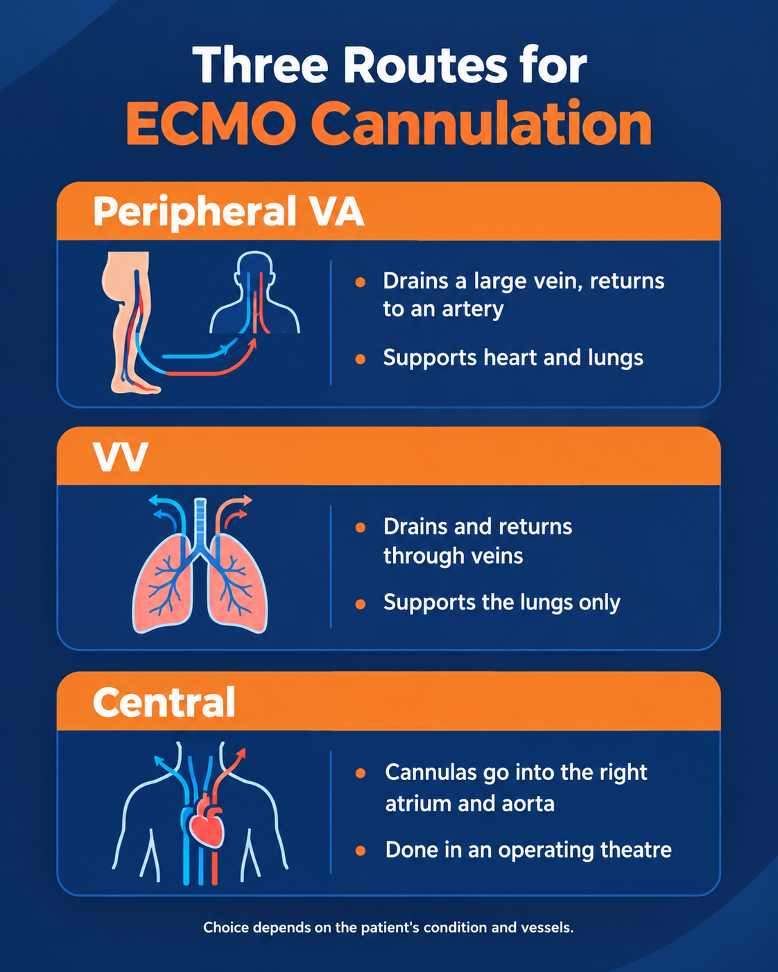
Before ECMO can start, blood needs to be drawn out of the patient’s body and then returned. That’s what cannulation does. Large tubes, known as cannulas, are placed in major blood vessels, and the blood is routed through an external heart lung machine on its way back. The position of those tubes matters a great deal, which is why imaging and careful checks are part of every ECMO cannulation.
According to Dr. Vishal Khullar, a Cardiac Surgeon in Mumbai, “The cannulas do the real work in ECMO. Choosing the right vessels and confirming the position matters as much as the machine itself.”
Cannula sites are chosen case by case, not from a fixed template.
Our ECMO therapy page explains how the treatment works and when it may be needed.
Have questions about ECMO treatment?
Strip away the jargon, and the circuit follows three simple steps: blood goes out, gets oxygenated, and returns.
|
Feature |
VA ECMO |
VV ECMO |
|
Organs supported |
Heart and lungs |
Lungs |
|
Blood drained from |
A large vein |
A large vein |
|
Blood returned to |
An artery |
A vein |
|
Typical sites |
Groin or neck vein, groin artery |
Groin and neck veins, or one neck tube |
|
Often chosen when |
The heart can’t pump enough, as in cardiogenic shock |
The lungs can’t oxygenate the blood |
How ECMO works: A pump draws blood out of the body and sends it through an oxygenator. That’s the part acting as an artificial lung: oxygen goes in, carbon dioxide comes out, and the blood returns to the patient. That gives the failing organ time to recover.
Breathing during ECMO: Some patients are awake and breathing for themselves, while others remain on a ventilator. Which of the two applies depends on the illness, and on how well the lungs still work.
Comfort and pain: Anesthesia or sedation is used while the cannulas go in. After that, pain relief continues for as long as the tubes are in place. Some soreness around the sites can happen.
How long it lasts: Usually days, not weeks. In a registry analysis of 2,699 adult VA ECMO runs, the median duration lasted 4 days and 41 percent of patients left hospital alive. What comes after depends far more on the illness behind it than on the machine.
If support has to last longer than ECMO allows, the ECMO vs LVAD comparison helps explain the available options.
Most peripheral cannulas are placed through the skin, so in this part of the ECMO procedure the main concern is the vessel itself. Then comes the checking.
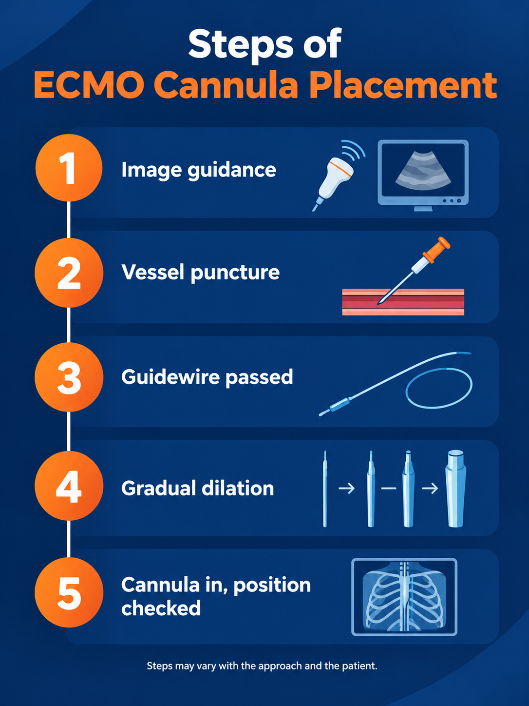
Imaging and puncture: First, ultrasound shows the size of the vessel and a safe place to enter. After that, a needle is passed into the vessel. This is how the Seldinger technique begins.
Guidewire and dilation: The needle then lets a thin guidewire through, and echocardiography or fluoroscopy can show its path. After that, dilators open the track a little at a time. Only then is the cannula pushed in.
Checking position: While the cannula is being advanced, echocardiography or fluoroscopy keeps track of the tips. A chest X ray afterwards shows where they ended up. What the team looks for on the film is the drainage tip near the right atrium. That’s the ECMO cannula position on X ray that counts.
Protecting the leg: A cannula sitting in the groin artery can starve the leg of blood. So the team often threads in a small extra catheter beyond it, called a distal perfusion catheter, and checks the limb regularly.
If the heart doesn’t recover, longer term choices are covered in our guide to heart replacement options.
ECMO cannulation is the process of drawing blood out of the body and returning it through the ECMO circuit. Where the cannulas go depends on whether the heart, the lungs or both need support. Most cases run from ultrasound to guidewire, gradual dilation and a final position check, and the leg gets extra attention when a leg artery is used. For a particular patient, the right approach is for a cardiac surgeon to decide.
Disclaimer: This blog is for educational and informational purposes only and should not be considered professional advice.

