Views: 19



The aorta is your body’s biggest artery. Its wall has three layers. An aortic intramural hematoma (IMH) means blood has seeped into the middle layer and stayed there. In an aortic dissection, the inner lining tears. In an IMH, there’s no classic tear. Doctors take it seriously. A weakened wall can tear or burst. IMH belongs to a group of conditions called acute aortic syndromes, which also includes aortic dissection and penetrating aortic ulcers. Two things guide treatment. First, where is it? In the ascending aorta (Stanford type A) or the descending aorta (Stanford type B)? And are complications present? The answers decide whether it needs urgent intervention or careful watching.
According to Dr. Vishal Khullar, a Cardiothoracic Surgeon in Mumbai, “Location matters a great deal. A hematoma in the ascending aorta, next to the heart, is more likely to rupture. Surgery is usually urgent. Further down, in the descending aorta, we often start with tight blood pressure control and repeat scans. The two behave quite differently, even though they look similar on a first scan.”
On a scan it looks like a thickened wall. You won’t see an obvious tear.
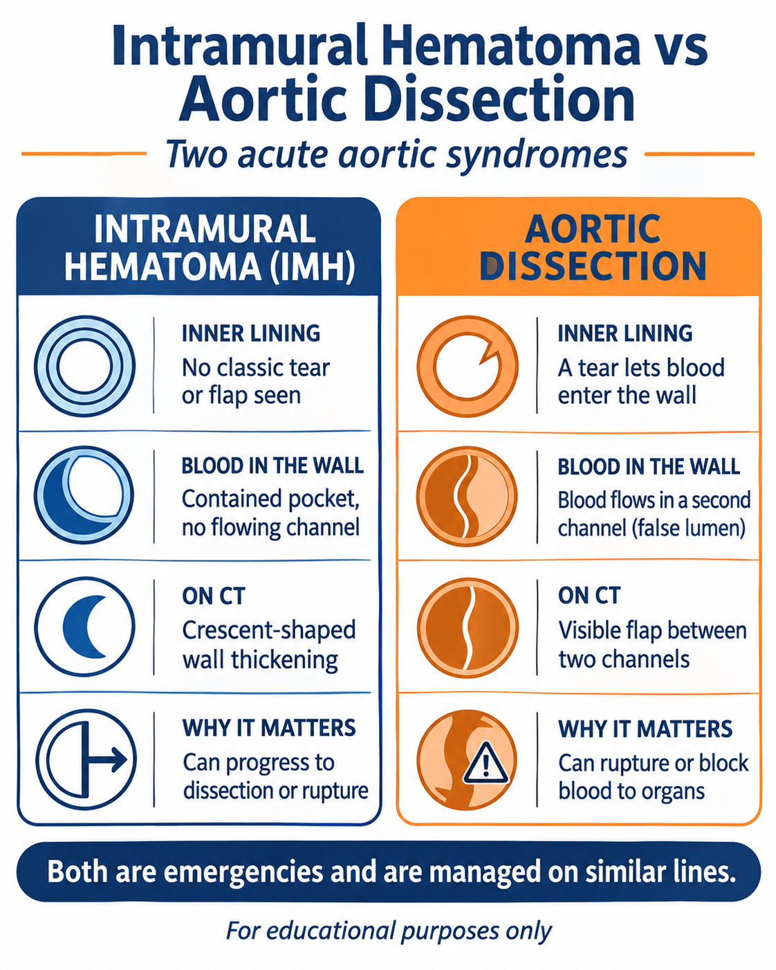
What it is: Blood pools inside the wall. On CT, that shows up as a crescent-shaped thickening. There is no flowing false channel as in dissection.
Common causes: Most cases trace back to years of high blood pressure. Atherosclerosis or a penetrating ulcer often plays a part. Chest injury and aneurysms are rarer triggers.
Symptoms to know: The pain comes on suddenly and it’s severe. Felt in the chest, it points to the ascending aorta. Felt in the back, the descending aorta.
Intramural hematoma vs dissection: A dissection starts with a tear that opens a second blood channel. A hematoma begins without either but can turn into one. Both are emergencies.
An IMH can turn into a dissection. That’s why its treatment borrows so much from aortic dissection surgery.
Why shouldn’t sudden, severe chest or back pain wait?
Intramural hematoma treatment comes down to location and complications. The first scan answers most of that.
|
Comparison |
Type A: ascending aorta |
Type B: descending aorta |
|
Where treatment starts |
Urgent surgery |
Medication and close monitoring |
|
Aim |
Prevent rupture and bleeding around the heart |
Keep pressure low, watch for change |
|
If a procedure is needed |
Open repair of the affected aorta |
TEVAR (stent graft) or open repair |
|
Early risk |
Higher |
Lower |
|
Afterwards |
Scans after repair |
Scans on a set schedule |
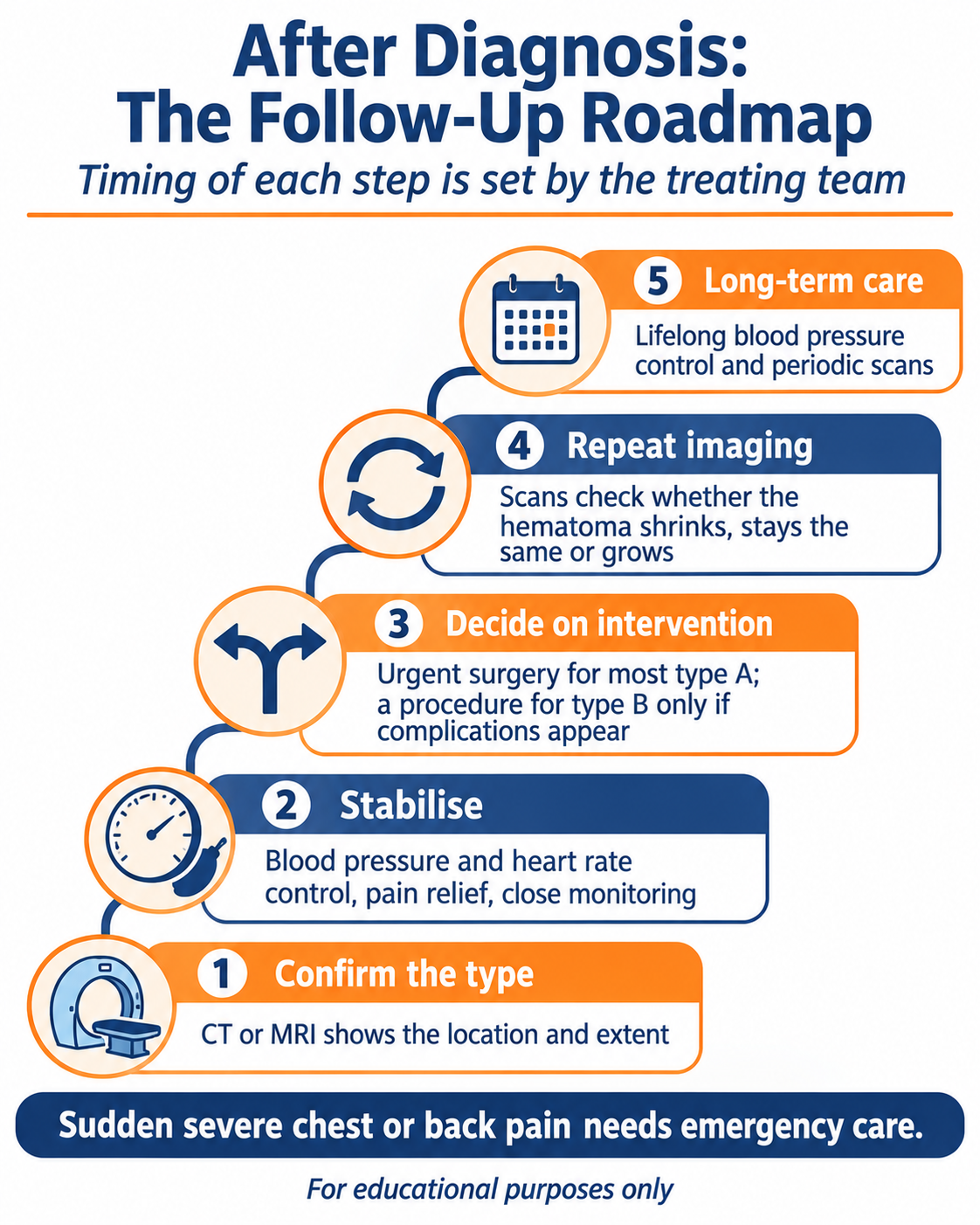
Type A: Guidelines say surgery shouldn’t wait. The damaged stretch of aorta is replaced. A few stable patients may begin with medicines and be watched very closely.
Type B: An intramural hematoma of the descending aorta usually starts with medicines that lower blood pressure and heart rate, plus pain relief. Scans follow to check progress.
When type B needs a procedure: Rupture, organs losing blood supply, pain that won’t settle, or growth on scans. The usual answer is TEVAR, a stent graft placed through the groin.
Abdominal aorta: An abdominal aortic intramural hematoma is rare, and research on it is limited. It is generally managed like type B disease, one case at a time.
If you’d like to know what recovery involves, this guide to care after aortic surgery walks through it.
The outlook depends on the type and on how early it’s found, and left alone a hematoma might shrink, stay put, or get worse without much warning. That uncertainty is the reason for repeat scans.
If it settles: Many hematomas shrink or hold steady, particularly in the descending aorta, when blood pressure is controlled and scans keep watch.
If it progresses: Some type B studies show 15 to 30% becoming a dissection. A thick hematoma, a wide aorta, or an ulcer-like bulge on the scan makes that likelier.
Prognosis by type: Type A is the riskier of the two early on. In-hospital deaths have been reported at about 40% for type A and under 10% for type B.
Long-term care: Blood pressure needs control for life, whichever treatment you’ve had. A beta blocker is often part of that. CT or MRI scans carry on at set intervals.
If repair is needed, it may be TEVAR or open surgery, and the aortic aneurysm surgery page explains both. If sudden, severe pain hits your chest or back, don’t wait it out: call 112.
An IMH is bleeding inside the aortic wall, and it’s handled a lot like a dissection. Aortic hematoma treatment follows location. For type A, that usually means urgent surgery. For type B, it means medicines and scans first. A procedure only comes into it if complications show up. Blood pressure control and regular scans carry on for life. Sudden, severe chest or back pain should always be treated as an emergency.
Disclaimer: This blog is for educational and informational purposes only and should not be considered professional advice.

