Trusted By
9,000+ Patients Globally
Experience of
30+ Years
7000+ Surgeries
Completed

Bicuspid Aortic Valve with Aneurysm

Bicuspid Aortic Valve Repair with Ascending Aortic Replacement and Endocarditis Management

A 52-year-old patient with severe shortness of breath, infective endocarditis, and a 50 mm ascending aortic aneurysm — treated by Dr. Vishal Khullar at Nanavati Max Super Speciality Hospital, Mumbai.

Patient Profile

The following clinical profile was prepared in line with Indian medical confidentiality guidelines. The patient consented to publication of his case for educational and clinical record purposes. His identity has been withheld.

Age

52 years

Gender

Male

Occupation

Working professional

City

Maharashtra (suburban region)

Presenting Complaint

Severe shortness of breath, reduced exertional tolerance

Diagnosis

Bicuspid aortic valve with ascending aortic aneurysm and infective endocarditis with aortic regurgitation

Duration of Issue

Progressive symptoms over several months prior to admission

Previous Treatments

Initial evaluation at a local hospital, including CT scan and echocardiography. No definitive surgical intervention prior to referral.

Date of Procedure

Recent surgical intervention

Outcome

Excellent — uneventful recovery, no post-operative complications

Patient identity withheld per confidentiality guidelines. Case published with documented consent.


The Problem

Clinical Condition

The patient was diagnosed with three overlapping cardiac conditions, each of which carried independent surgical urgency. The combination made the case unusually complex.

The first finding was a bicuspid aortic valve, a congenital condition in which the valve has only two leaflets instead of the usual three. This abnormal structure had been present since birth, and over decades it had begun to fail mechanically. The second finding was an ascending aortic aneurysm measuring 50 millimetres, well above the 45 to 50 mm surgical threshold recommended by current cardiothoracic guidelines. Earlier imaging at the patient’s local hospital had estimated the aneurysm at 28 mm, indicating rapid progression. The third finding, and the most acute, was infective endocarditis affecting the aortic valve. Bacterial vegetations had destroyed parts of the leaflet tissue and the valve had begun to leak severely. Blood was flowing backward into the heart with every contraction.

Emotional and Psychological Impact

By the time the patient reached Mumbai, his breathlessness had become severe enough to limit ordinary movement around the house. The family described a period of considerable anxiety. They had visited a local hospital first, where a CT scan and echocardiography revealed the aortic leak. A relative referred them to Nanavati Hospital and to Dr. Vishal Khullar.

The family arrived without a clear understanding of what surgery would involve or what the recovery would look like. The diagnosis itself was difficult to accept, given that the patient had appeared otherwise healthy until the breathlessness began. The clinical team focused early consultations on patient and family education, recognising that informed consent for a procedure of this magnitude required time and clarity.

Consultation and Treatment Plan

Pre-Surgical Assessment by Dr. Vishal Khullar

The patient underwent a complete cardiac and aortic workup before any surgical decision was made. The assessment was structured to clarify the severity of each condition and to establish whether a single combined operation was both feasible and safer than staged procedures.

  • Transthoracic echocardiography to grade the severity of aortic regurgitation and to confirm the bicuspid morphology of the native valve.
  • CT aortogram to measure the ascending aortic aneurysm and to map the aortic arch anatomy. Imaging at Nanavati Hospital documented the aneurysm at 50 mm, larger than the patient’s earlier scan suggested.
  • Blood cultures and infection markers to confirm infective endocarditis and to identify the causative organism so antibiotic therapy could be directed appropriately.
  • Coronary angiography to rule out concurrent coronary artery disease that might require simultaneous bypass grafting.
  • Anaesthetic and renal fitness review, since the planned procedure involved deep hypothermic circulatory arrest and prolonged cardiopulmonary bypass.

Why a Combined Procedure Was Chosen

Several treatment paths were considered. Dr. Khullar’s team weighed the risks of staged surgery against a single combined operation. The combined approach was selected for the following clinical reasons.

  • The aneurysm size of 50 mm carried a real risk of dissection or rupture. Delaying its repair to allow recovery from valve surgery would have left the patient in a high-risk window for several weeks.
  • Active infective endocarditis required removal of the diseased valve along with any infected tissue surrounding it. Operating on the valve without simultaneously addressing the dilated aorta would have meant returning the patient for a second sternotomy within months, with significantly higher cumulative risk.
  • A bicuspid valve combined with aortopathy is a recognised pattern in cardiothoracic surgery. Established consensus guidelines (American Association for Thoracic Surgery, 2018) recommend simultaneous repair of the valve and ascending aorta when the aneurysm exceeds 45 mm in patients undergoing valve surgery.
  • The patient had adequate cardiac reserve and good organ function. He was a suitable candidate for the longer operating time and the period of circulatory arrest that the combined approach demanded.

The plan was finalised after a multidisciplinary discussion involving cardiac anaesthesia, perfusion, and intensive care teams. The patient and his family were counselled in detail about the planned circulatory arrest, the use of a vascular graft, and the prosthetic valve options.

Pre-Operative Imaging and Anatomy

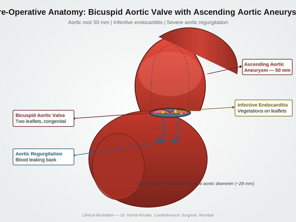
Imaging confirmed the diagnostic triad. The illustration below summarises the anatomical findings observed on transthoracic echocardiography and CT aortogram.

Picture1

Figure 1. Pre-operative anatomy showing bicuspid aortic valve with infective vegetations, ascending aortic aneurysm at 50 mm, and severe aortic regurgitation.

Procedure Details

Step-by-Step Surgical Overview

  • Median sternotomy was performed to access the heart and the ascending aorta.
  • The patient was placed on cardiopulmonary bypass. Arterial cannulation was sited to allow selective antegrade cerebral perfusion during the period of circulatory arrest.
  • Systemic cooling was initiated. The body was brought down to deep hypothermia to protect the brain and visceral organs against ischaemia.
  • The aortic cross-clamp was applied. Cardioplegic solution was administered to arrest the heart in diastole.
  • The diseased ascending aorta was opened. The bicuspid aortic valve was inspected and the infected leaflets and vegetations were excised. All visibly infected tissue was debrided to reduce the risk of recurrent endocarditis.
  • Circulatory arrest: Body circulation was stopped for 24 minutes to allow safe excision of the dilated aortic segment. The brain and visceral organs are highly vulnerable to ischaemia during this window. Selective cerebral perfusion was maintained to protect neurological function throughout.
  • A Dacron vascular graft was sutured into place to replace the aneurysmal segment of the ascending aorta. Distal and proximal anastomoses were tested for haemostatic integrity.
  • A mechanical aortic valve prosthesis was implanted at the aortic annulus. The valve was tested intraoperatively for free leaflet motion and competent closure.
  • Rewarming was initiated. Cardiopulmonary bypass was gradually weaned, and the heart was restarted in sinus rhythm.
  • The chest was closed in layers with stainless steel sternal wires and absorbable suture material. Drains were positioned for post-operative monitoring.

Procedure Facts

Duration

Approximately 6 to 7 hours

Anaesthesia

General anaesthesia with invasive haemodynamic monitoring

Implants Used

Mechanical bileaflet aortic valve prosthesis and Dacron ascending aortic graft

Approach

Median sternotomy with cardiopulmonary bypass

Circulatory Arrest

24 minutes with selective antegrade cerebral perfusion

Intraoperative Complications

None

Hospital Stay

Three days in ICU followed by ward recovery

Picture2

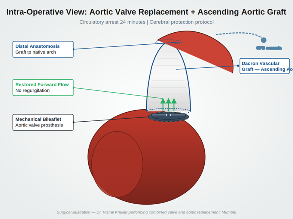
Figure 2. Intraoperative reconstruction. The ascending aorta has been replaced with a Dacron graft and the bicuspid aortic valve has been replaced with a mechanical bileaflet prosthesis.

Post-Operative Results

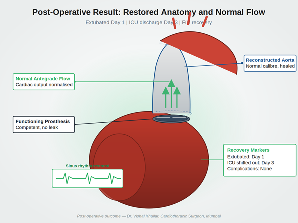
The combined procedure achieved its three primary objectives. The infected aortic valve was completely excised and replaced with a competent prosthesis. The dilated ascending aorta was replaced with a vascular graft, removing the risk of dissection. Forward blood flow from the left ventricle to the systemic circulation was restored. Aortic regurgitation was eliminated.

Post-operative recovery was uneventful. The patient was extubated on post-operative day one. He was shifted out of the ICU on post-operative day three. There were no surgical, neurological, or infective complications.

Picture3

Figure 3. Post-operative result. Reconstructed aortic anatomy with normal calibre, functioning prosthetic valve, and restored sinus rhythm.

Outcomes at a Glance

Outcome Metric

Result

Symmetry and Cardiac Function

✓  Excellent. Sinus rhythm restored on weaning from bypass.

Surgical Outcome

✓  Complete excision of infected tissue and aneurysmal segment.

Patient Recovery

✓  Smooth and uncomplicated. Family confirmed full symptom resolution.

Complications

✓  None during ICU stay or ward recovery.

Recovery Trajectory

✓  On track. Discharged within the expected timeframe.

Patient and Family Feedback

The following feedback was recorded during the patient’s clinical follow-up. It has been transcribed from the family’s original words. Minor language corrections have been made for readability while preserving the substance of what was said. A video recording of the patient’s family and Dr. Vishal Khullar narrating the case is included below.

My son had been suffering for some time. We took him to a local hospital first. They did a CT scan and a sonography. After the sonography, they told us there was a leak in his heart valve. We were worried. A relative gave us a letter for Nanavati Hospital and told us to consult Dr. Vishal Khullar. We met the doctor. He explained that surgery was needed. He said there was no other option. The aneurysm had grown to 50 millimetres. After the operation, my son recovered completely. We are grateful to the doctor and his team.

— Father of the patient — recorded during follow-up review

Watch the patient’s family and Dr. Vishal Khullar describe the case in their own words:

▶  WATCH THE FULL CASE NARRATION

Bicuspid Aortic Valve with Ascending Aortic Aneurysm and Infective Endocarditis — Patient Family and Dr. Vishal Khullar

Surgeon’s clinical note on the case:
The patient was a 52-year-old male who presented with severe shortness of breath. He was diagnosed with a bicuspid aortic valve, ascending aortic aneurysm, and infective endocarditis of the aortic valve. The infection had caused the valve to leak. Blood was flowing back into the heart with each contraction. To replace the diseased aortic segment, we had to stop body circulation for 24 minutes. This is a high-risk operation because the brain and several internal organs are vulnerable to ischaemia, and their function must be actively protected. After surgery the patient did well. He was extubated on post-operative day one. There were no complications. He was shifted out of the ICU on post-operative day three.
— Dr. Vishal Khullar, Cardiothoracic Surgeon, Nanavati Max Super Speciality Hospital, Mumbai

POST-PROCEDURE CARE AND RECOVERY

Discharge Instructions Given to the Patient

  • Sternal precautions for six weeks. No lifting of weights heavier than two to three kilograms.
  • Lifelong anticoagulation with warfarin for the mechanical aortic valve. INR target range was specified, with weekly monitoring during the first month.
  • A six-week course of intravenous antibiotics directed against the organism cultured at admission, transitioned to oral therapy as appropriate.
  • Wound care for the sternal incision, with strict instructions to report fever, redness, drainage, or chest wall instability.
  • Cardiac rehabilitation referral, beginning with supervised walking and progressing as tolerated.
  • Scheduled follow-up at two weeks, six weeks, three months, and six months post-discharge, with echocardiography at the six-week and six-month visits.
  • Dental review and prophylactic antibiotic guidance for any future dental work, given the prior endocarditis history.

Recovery Timeline

Timeframe

What the Patient Could Expect

Day 1 to 3

ICU recovery. Extubation on day one. Haemodynamic stabilisation. Transition to ward by day three.

Week 1 to 2

Ward recovery. Anticoagulation started and titrated. Antibiotic regimen continued. Sutures and drains removed in stages.

Week 4 to 6

Light activity at home. Cardiac rehabilitation initiated. First post-discharge follow-up at week two, then week six with echocardiography.

Month 3

Return to most non-strenuous routine activities. Sternal healing complete. INR stable on home monitoring.

Month 6

Six-month review with echocardiography. Prosthesis function confirmed. Patient discharged from active surgical follow-up to long-term cardiology care.

FAQs

Q1. What is a bicuspid aortic valve and why does it need surgery?

A bicuspid aortic valve is a congenital condition in which the valve forms with only two leaflets instead of the usual three. Many people live with it for decades without symptoms. Over time the valve can become stiff, leak, or develop infections, and it is also linked to enlargement of the ascending aorta. When complications develop, surgical repair or replacement is needed. Dr. Vishal Khullar performs bicuspid aortic valve surgery at Nanavati Max Super Speciality Hospital in Mumbai.

Q2. Why was both the valve and the aorta replaced in the same operation?

When a bicuspid valve is associated with an ascending aortic aneurysm above 45 to 50 millimetres, current cardiothoracic guidelines recommend repairing both at the same time. This avoids a second high-risk surgery within months and addresses the dissection risk before it can rupture. In this case the aneurysm measured 50 millimetres and the valve was acutely infected, which made a single combined procedure the safer route.

Q3. What is circulatory arrest and how is it carried out safely?

Circulatory arrest is a controlled period during open aortic surgery in which body circulation is briefly stopped. It allows the surgeon to operate on a bloodless aortic field. Patient safety depends on deep cooling and on selective perfusion of the brain. In this case, body circulation was stopped for 24 minutes while cerebral perfusion was preserved. The technique is high-risk and is performed at centres with experienced aortic teams.

Q4. How long does recovery take after combined valve and aortic graft surgery in Mumbai?

Most patients spend two to three days in the ICU and another seven to ten days in the ward. Sternal healing takes around six weeks. Light routine activity returns within two to three months, and full recovery with cardiac rehabilitation is usually complete by the six-month mark. Patients on a mechanical valve continue lifelong anticoagulation under medical supervision. Specific timelines can vary, and the cardiac team at Nanavati Max Super Speciality Hospital in Mumbai sets follow-up schedules to suit each case.

Q5. Why choose Dr. Vishal Khullar for complex aortic and valve surgery?

Dr. Vishal Khullar is the Director of Cardiovascular and Thoracic Surgery, Heart and Lung Transplantation, and Mechanical Circulatory Support at Nanavati Max Super Speciality Hospital in Mumbai. He trained and held senior positions at Mayo Clinic in Rochester and Cleveland Clinic in Ohio. His surgical experience covers more than 7,000 cardiac operations, including complex aortic procedures, infective endocarditis, redo cardiac surgery, and heart and lung transplantation. He works with a multidisciplinary aortic team, which is the appropriate setting for cases of this complexity.


Book Online Consultation Book An Appointment